
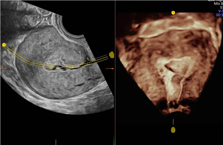
Infertility Sonography
Ultrasound can contribute to fertility assessment by examining pelvic anatomy, follicles and the uterine lining.
Information from a scan
The doctor may assess the uterus, ovaries and developing follicles. Scanning can identify some structural concerns and support planning for selected fertility treatments.
Timing the examination
The timing may depend on your menstrual cycle and the purpose of the scan. Follicular monitoring can involve more than one visit; follow the schedule provided by your treating team.
Part of a wider assessment
A scan alone cannot explain every fertility concern or establish whether pregnancy will occur. Blood tests, semen analysis or other investigations may be considered alongside ultrasound findings.
This information supports a discussion with your doctor. Suitability, availability and any specialist referral should be confirmed with the hospital. Individual care plans and outcomes vary.
All sonography topics